

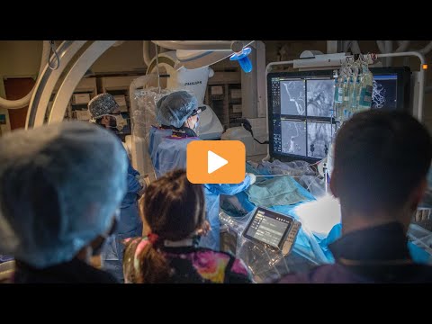
In front of a dozen international observers, St. Michael’s Hospital’s neurovascular team carried out yet another world-first: advanced, intravascular imaging of a man previously treated for the narrowing of arteries inside the brain, which causes strokes.
For months, Dr. Vitor Pereira, a neurosurgeon, global expert in minimally-invasive procedures involving brain and spinal cord blood vessels, and head of St. Michael’s RADIS lab, has been testing a new neuroimaging device on silicon models. The Vis-M probe is the latest in optical coherence tomography (OCT) technology, first used in the field of ophthalmology 25 years ago. It’s been adapted to get a better view of the brain vessel during an angiogram, for example.
OCT systems use glass fibre optics to create extraordinarily high-resolution, cross-sectional images of tissues. When the Vis-M probe is placed at the end of a catheter, the resolution is 10 times current imaging technologies. That means clinicians can see micron-sized particles in ways they never could before.
When Dr. Pereira and his team detected a narrowing of the blood vessel inside an 82-year-old patient’s brain several months after his last surgery, they wondered if he had atherosclerosis, a build-up of plaque on the artery walls, or even a blood clot. Using the Vis-M probe inside his brain vessel told the physicians how to adjust the patient’s treatment to prevent more procedures.
Currently, St. Michael’s is the only hospital in the world to offer the OCT procedure.
“This technology is not only revolutionizing medical imaging, but also transforming our understanding of strokes and other vascular diseases from brain vessels,” says Dr. Pereira, who holds the Schroeder Chair in Advanced Neurovascular Interventions at the Schroeder BRAIN&HEART Centre. “We can see the arterial wall better to assess complex brain aneurysms, examine the narrowing of arteries that leads to stroke and evaluate intracranial stenting surgeries. We can increase the precision and safety of our procedures, and improve patient outcomes.”
Nicole Cancelliere, the neurovascular research program manager, radiographer and co-lead of the RADIS lab, says this application of the Vis-M probe represents another one of its ‘benchtop to bedside’ translations. The RADIS team is known for its world-firsts, including the first robotic-assisted neurovascular intervention in 2019.
“We're committed to being at the forefront of innovation, partnering to develop intracranial devices, such as stents, remote robotic neurosurgery and now advanced intravascular brain imaging, which gives us eyes on the inside of patients’ brains,” Cancelliere says.
It’s based on years of international research and development, Dr. Pereira acknowledges.
“It was an honour to lead this worldwide effort,” he says. “Together with Gentuity, the Vis-M probe’s manufacturer, the University of Massachusetts, which performed the pre-clinical work, Buenos Aires’ ENERI Institute and global health centres, we were able to translate this technology for our patients.”
Next up, St. Michael’s will train hospital centres in Argentina and other countries on how to use the Vis-M probe, so they can deliver treatments for cerebrovascular diseases to their own patients.
Meet the team behind this exciting medical innovation. Watch now.
Donate to St. Michael's Hospital Foundation.


